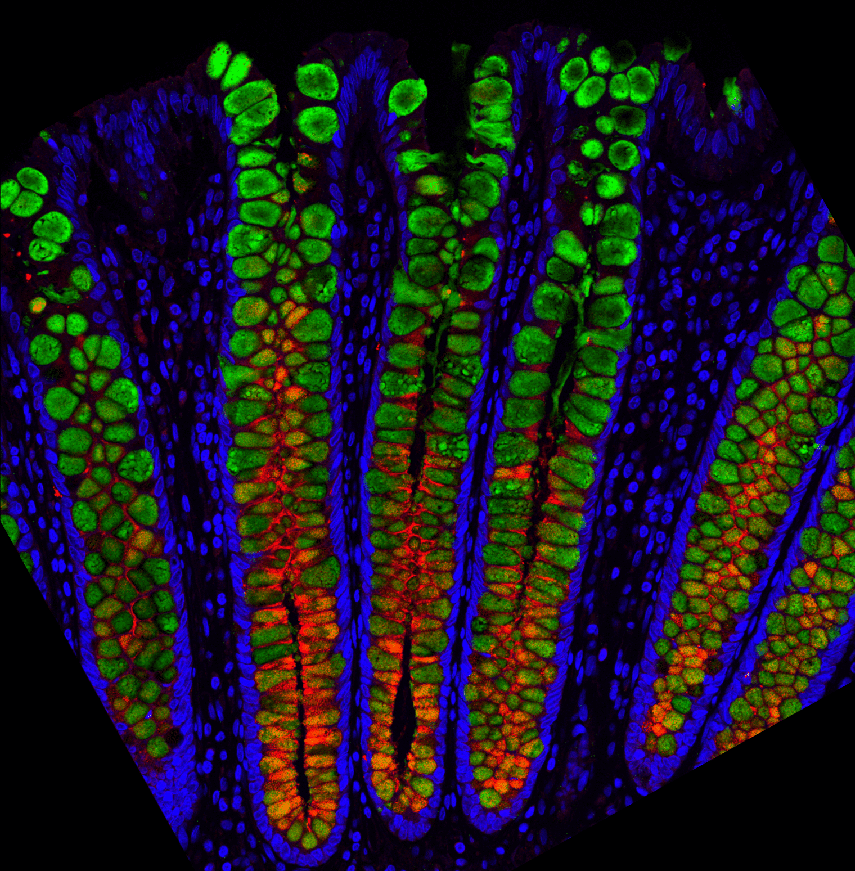
For an area involved in almost every aspect of human health, scientists still have much to learn when it comes to the gut. Now, for the first time, researchers from the University of North Carolina Health Care have successfully mapped out the entire human GI tract, including how different cells living in the gut are important to daily life.
“Our lab showed it’s possible to learn about each cell type’s function in important processes, such as nutrient absorption, protection from parasites, and the production of mucus and hormones that regulate eating behavior and gut motility,” says Scott Magness, PhD, associate professor in the Joint UNC-NC State Department of Biomedical Engineering and senior author of the study, in a statement. “We also learned how the gut lining might interact with the environment through receptors and sensors, and how drugs could interact with different cell types.
‘We want a ground level view to know as much as possible’
One of the research areas of interest was uncovering what cell types are involved in unpleasant side effects such as vomiting and internal bleeding after taking medications. They focused their attention on a single-cell thick layer called the epithelium that separates the intestines and colon from the rest of the body. Before this study, researchers could only take small samples — about the size of a grain of rice — from the digestive tract to study the epithelium.
“Such exploration would be like looking at the United States from space but only investigating what’s going on in Massachusetts, Oklahoma, and California,” Dr. Magness says. “To really learn about the country, we’d want to see everything.”
The rice-sized samples would then have to be broken down into smaller pieces to identify all epithelial cell types and their features. However, the current study used a different technique of sampling thousands of individual cells from the small intestine and colon to create a map of the GI tract. The map would then be used to predict the potential role of the cells based on the gene expression of nearby cells.
“Not only do we want to identify where the cells are located, but we want to know exactly which cell types do what, and why,” Joseph Burclaff, PhD, co-first author of the paper also said in the press release. “So, staying with the map analogy, we don’t want to just say, ‘oh, there’s North Carolina’. We want to know where to get the best barbecue. We want a ground level view to know as much as possible.”
How did scientists develop the gut map?
To create the map to study cell gene expression, the research team studied the GI tracts from three organ donors. The epithelial layer was removed from the donated tracts and exposed to enzymes to break it down to individual cells. RNA sequencing technology would then be used to study the gene expression of each cell and the amount it was expressing.
“The picture we get from each cell is a mosaic of all the different types of genes the cells make and this complement of genes creates a ‘signature’ to tell us what kind of cell it is and potentially what it is doing,” explains Dr. Magness. “Is it a stem cell or a mucous cell or a hormone-producing cell or an immune-signaling cell?
Dr. Burclaff adds: “We were able to see the differences in cell types throughout the entire digestive tracts, and we can see different gene expression levels in the same cell types from three different people. We can see the different sets of genes turned on or off in individual cells. This is how, for instance, we might begin to understand why some people form toxicity to certain foods or drugs and some people don’t.”
The study is published in the journal Cellular and Molecular Gastroenterology and Hepatology.

Hopefully steps closer to curing crohns
It would be amazing if this study leads to a cure for Crohn’s disease!
This research is amazing and is going to facilitate faster insight into curing gastrointestinal disease across the spectrum.
My you all suceed in your efforts.
Friends of mine suffer from this.
Godspeed.